K5 Polysaccharides Inhibit SARS-CoV-2 Infection by Preventing Spike-Proteolytic

Author(s)

M. Milanesi, C. Urbinati, L. Zimmermann, P. Oreste, A. Zani, A. Caruso, F. Caccuri, V. Laketa, P. Chlanda, R.C. Wade, M. Rusnati1 & G. Paiardi

Sources

Milanesi, M., Urbinati, C., Zimmermann, L. et al. K5 polysaccharides inhibit SARS-CoV-2 infection by preventing spike-proteolytic priming. npj Viruses 4, 3 (2026). https://doi.org/10.1038/s44298-025-00163-4

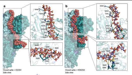
PSARS-CoV-2 spike glycoprotein is a promising drug target due to its crucial role in viral infection.Heparin, a long linear polysaccharide that inhibits SARS-CoV-2 infection by acting on spike, has limited antiviral applications due to its anticoagulant effect. E. coli K5 polysaccharides share the same structure as the heparin precursor and can be chemically modified to be devoid of anticoagulant activity. Here, biochemical assays and computer simulations reveal that K5 with high degree of sulfation at O- (K5OSH) or N- and O-positions (K5NOSH) bind spike with higher affinity than heparin, preventing its binding to ACE2 and furin cleavage. This mechanism is supported by cell syncytia assays showing that K5OSH and K5NOSH inhibit viral infection by blocking membrane fusion. Infection assays for SARS-CoV-2 Wuhan-Hu-1 and Omicron BA.1 variants corroborate their antiviral activity. These results support the therapeutic potential ofK5OSHandK5NOSHagainstSARS-CoV-2, with K5OSH displaying more promising activity profile.riming

 MD simulations of K5OSH and K5NOSH on the closed spike conformation reveal tighter packing of the spike subunits when K5OSH rather than heparin is bound. a, b Representative structures obtained after MD simulation of the spike bound to three K5OSH a or K5NOSH b chains. The SA, SB, and SC spike subunits are shown as molecular surfaces in cyan, teal, and blue, respectively. The 31mer K5 chains that span fromtheRBDof one spike to the S1/S2 site of the adjacent subunit are shown in van der Waals sphere representation colored by element with carbons in pink and purple for K5OSH and K5NOSH, respectively. Insets show the interactions of the K5 chains with residues in the down-RBD (top) and S1/S2 sites (bottom). The spike and its interacting residues are depicted in cartoon and stick representations, respectively, and colored according to the respective subunit.  H-bonds are shown as yellow dashed lines. N-glycans covalently attached to the spike are not shown for clarity.

Latest news

Neonatal sepsis, a systemic infection that occurs in infants within their first 28 days, is...

Chirality has become a key design principle for the construction of peptide materials. In contrast,...

Sialic acids are the terminal monosaccharides of the glycocalyx that play a crucial role in...

Glycans are branched, structurally diverse, and highly flexible biomolecules. These characteristics make glycoanalytics and structural...