
Laser microdissection-assisted lectin microarray provides quantitative and qualitative information on glycans on proteins expressed in microscopic regions of formalin-fixed paraffin-embedded tissue sections. For the effective visualization of this “tissue glycome mapping” data, a novel online tool, LM-GlycomeAtlas (https://glycosmos.org/lm_glycomeatlas/index), was launched in the freely available glycoscience portal, the GlyCosmos Portal (https://glycosmos.org). In LM-GlycomeAtlas Version 1.0, nine tissues from normal mice were used to provide one data set of glycomic profiles.
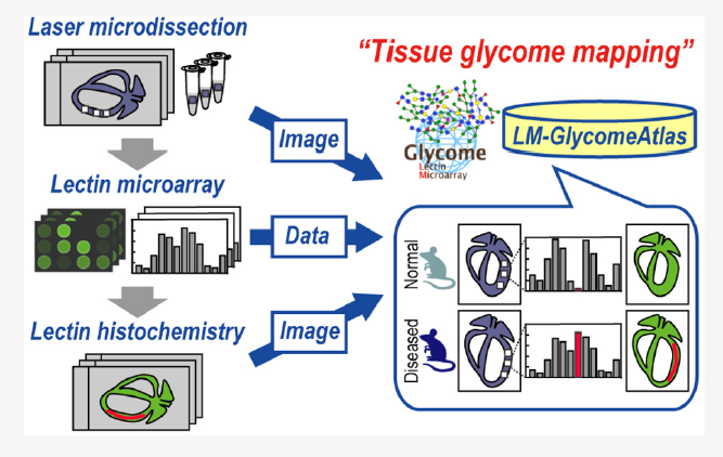
An updated version of LM-GlycomeAtlas allows the users to display multiple histological images of interest (e.g., diseased and normal mice), thereby facilitating the evaluation of tissue glycomic profiling and glyco-pathological analysis. The use of such updated interfaces allowed visualisation of 451 glycomic profiling data and 42 histological images obtained from 14 tissues of normal and diseased mice. Throughout an easy integration with other tools for glycoproteomic data and protein glycosylation machinery, LM-GlycomeAtlas stands as a valuable open resource that contributes to both glycoscience and proteomics communities.