Glycosphingolipids are among the most structurally complex lipids in mammalian membranes. Comprising a ceramide backbone and a glycan headgroup, they present the combined descriptive challenges of both lipids and carbohydrates. Their carbohydrate moiety, characterized by high conformational flexibility, serves as a receptor for carbohydrate-binding proteins, including lectins, such as those of pathogenic origin. Glycosphingolipids also exhibit heterogeneous lateral diffusion, thereby promoting the formation of nanoscale functional complexes during signal transduction. In addition to glycosphingolipids, these complexes may contain GPI-anchored proteins, transmembrane proteins, and phospholipids from both membrane leaflets, the latter connected through acyl-chain interdigitation, thereby enabling signal transmission from the extracellular environment to the cell interior.
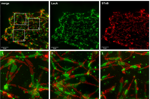
The formation of such molecular assemblies spans a broad range of spatial and temporal scales and remains largely unresolved because of the limited resolution of current observational techniques. In this review, the authors integrate insights into glycosphingolipid dynamics in membranes and their interactions with carbohydrate-binding proteins, drawing on high- and super-resolution fluorescence microscopy as well as molecular modeling. Using examples including the gangliosides GM1 and GM3 and the globoside globotriaosylceramide (Gb3, also known as CD77 or Pk antigen), the authors illustrate how the gap in spatial and temporal resolution between experimental and computational approaches is steadily narrowing. The selected case studies encompass investigations in both cellular and membrane model systems, including giant unilamellar vesicles and supported lipid bilayers, as well as in silico lipid bilayers. By reducing environmental complexity, these systems help identify the key molecular determinants underlying functional complex formation, membrane bending, endocytosis, and signaling.