The pectin puzzle: Decoding the fine structure of rhamnogalacturonan-I (RG-I) in Arabidopsis thaliana uncovers new pectin features

Author(s)

L. Zhang, J.Vlach, I.M. Black, S. Archer-Hartmann, C. Heiss, P. Azadi & B. R. Urbanowicz

Sources

Carbohydrate Polymers, 368, Part 2, (2025) 124161 https://doi.org/10.1016/j.carbpol.2025.124161

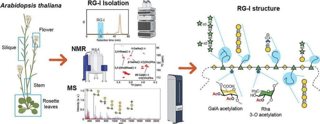
Pectin can be divided into four distinct structural categories: homogalacturonan, xylogalacturonan, rhamnogalacturonan I (RG-I), and rhamnogalacturonan II. While the structural diversity of homogalacturonan, xylogalacturonan, and rhamnogalacturonan II is well understood, the structural features of RG-I remain unclear. In this study, we used a variety of analytical techniques to conduct a detailed structural analysis of RG-I in the model species Arabidopsis thaliana. Starting with highly purified RG-I extracted from various Arabidopsis tissues, we used comparative linkage analysis, nuclear magnetic resonance spectroscopy, and mass spectrometry to analyze enzymatically digested RG-I oligosaccharides. In addition to the presence of the canonical α-1,5-arabinan, β-1,4-galactan, β-1,6-galactan, and arabinogalactan RG-I side chains of varying lengths, our results demonstrate that a significant proportion of the β-1,6-galactan is terminated by either 4-O-methyl β-glucuronic acid (GlcA) residues or, to a lesser extent, by β-GlcA lacking the Me-ether group. Notably, O-acetylation of RG-I GalA residues is a minor modification, with only 10% of the backbone Rha residues being 3-O-acetylated. Furthermore, most of the acetylated Rha residues are additionally branched with β-galactose substituents. Taken together, the combined results of these different analytical techniques represent the most comprehensive structural overview of Arabidopsis thaliana RG-I to date.

Figure schematic representation of RG-I structures identified in Arabidopsis. The RG-I backbone is formed by a disaccharide repeat of →2)-α-L-Rha(1→4)-α-D-GalA(1→. Rha residues can be substituted at O-4 with linear or branched arabinan, galactan, or arabinogalactan side chains. Our data indicate α-1,3-arabinan side chains are further substituted with single Araf residues in the 2-, 3-, and 2,3-positions. Arabinans were directly attached to the backbone, but the type of linkage is unknown. β-1,4 and β-1,6 linked galactan side chains were observed. β-1,6 linked galactans were substituted with single α-1,3-linked Araf residues. A portion of the galactan side chains was terminated by GlcA residues that carried a methyl-ether at the O-4 position. Arabinogalactans were observed, but we were unable to determine how they are attached to the backbone, and may be via Gal or an Ara residue. The type of linkage to the RG-I backbone could be resolved by NMR only for the single-Gal side chain (β-1,4) but not the other side chains. Acetylation of backbone Rha residues was the major acetyl modification of RG-I found in this study. Mono- and di-acetylated GalA residues were also observed. Terminal β-linked Araf residues (in the magenta dash box) were identified, but their point of attachment is unknown. Black lines: simplified RG-I backbone. Black dashed lines: the chains can be longer and/or substituted. Magenta lines: linkages not deciphered in this study. Magenta dashed lines: linkages not deciphered, and the chains can have more than one Gal or Araf.

Latest news

The gastrointestinal (GI) tract is home to trillions of microorganisms that live in symbiosis with...

Oligosaccharyltransferase (OST) catalyses the key step of N-glycosylation, transferring immature N-glycans to select Asn...

Glycans are complex carbohydrates that exhibit extraordinary structural complexity and stereochemical diversity while playing essential...

Efforts to fully understand the structure-to-function relationships of glycosaminoglycans (GAGs) have been hampered by the...