Revealing structure and shaping priorities in plant and fungal cell wall architecture via solid-state NMR

Author(s)

Xiao P, Sahu P, Pfaff SA, Ankur A, Ranasinghe YK, Gow NAR, Latgé JP, Cosgrove DJ, Wang T.

Sources

Revealing structure and shaping priorities in plant and fungal cell wall architecture via solid-state NMR. Cell Surf. 2025 Oct 31;14:100159. doi: 10.1016/j.tcsw.2025.100159. PMID: 41230029; PMCID: PMC12603724.

Plant and fungal cell walls play essential roles in growth, adaptation, and survival, with their intricate structures influencing resistance to stress and susceptibility to antifungal or biomass-degrading strategies. Understanding how these walls form, remodel, and function at the molecular level is crucial for both medical and biotechnological applications. Solid-state nuclear magnetic resonance (ssNMR) has emerged as a uniquely effective method for this, revealing the structure, dynamics, and interactions of intact biopolymers without disturbing their natural organization. Recent studies demonstrate how this technique can be highly effective. Variations in structure, polymer interactions, and species-specific remodeling affect mechanical strength, drug resistance, and stress responses. Applications include examining lignin-carbohydrate packing during plant stem development, observing changes in fungal walls when treated with wall-targeting antifungals such as echinocandins and nikkomycins, and analyzing the functional array of glucans, chitins, and mannans. These findings unveil conserved principles of polymer assembly across different kingdoms and open new possibilities for antifungal development and biomass utilization. Ongoing advancements in sensitivity and resolution are expected to expand ssNMR capabilities and enhance its role in linking structural diversity to biosynthetic complexity and biological function.
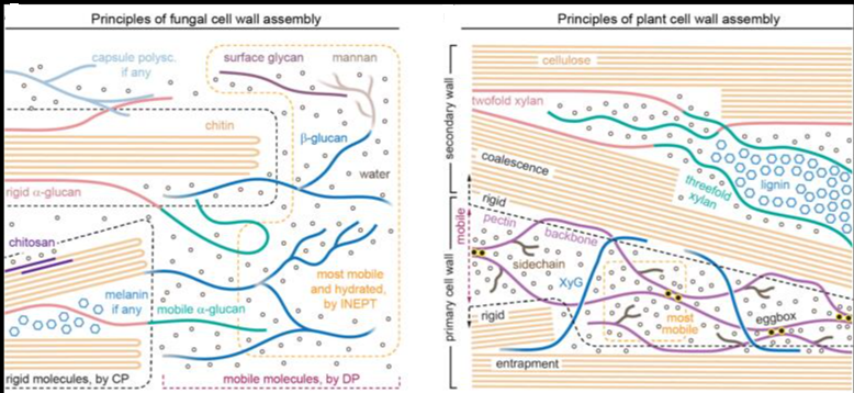
Principles of fungal and plant cell wall assembly selectively visualized by ssNMR. The structural principles governing polymer packing and supramolecular assembly, as revealed by ssNMR, are illustrated for (A) fungal cell walls and (B) plant primary and secondary cell walls. For chitin and cellulose, the number of strands depicted are only spacer holders and does not correspond to the actual number of chains within the microfibrils. In the fungal cell wall illustration, key ssNMR experiments highlight selective detection of rigid molecules (via CP), mobile molecules (via DP with short recycle delays), and highly mobile fractions (via INEPT), enclosed in dashed lines in black, purple, and yellow, respectively. In the plant cell wall illustration, dashed lines are used to separate three domains: highly mobile regions (yellow, mostly pectin sidechains and backbones), mobile regions (purple mostly matrix polysaccharides), and rigid regions (black; cellulose microfibrils and associated matrix polysaccharides in primary cell walls, and most molecules in secondary cell walls).

Latest news

This study uncovers a surprising, non-linear behavior in how polysaccharides change shape when you add...

Isotopic metabolomics reveals that plant species exhibit highly divergent carbon allocation and metabolic rewiring strategies...

The Protein Data Bank (PDB), established in 1971, is the primary global, open-access archive for...

Carbohydrates are among the most abundant and structurally diverse biomolecules in nature, playing central roles...