Ångström-resolution imaging of cell-surface glycans

Author(s)

L.A. Masullo, K. Almahayni, I. Pachmayr, M. Honsa, L. Heinze, S. Fritsche, H. Grabmay, R. Jungmann, & L. Möckl

Sources

bioRxiv preprint doi: https://doi.org/10.1101/2025.02.07.637003

Glycobiology is rooted in the study of monosaccharides, Angstrom-sized molecules that are the building blocks of intricate glycosylation patterns. Glycosylated biomolecules form the glycocalyx, a dense coat encasing every human cell with central relevance – among others – in immunology, oncology, and virology. In order to understand glycosylation function, visualizing its molecular structure is fundamental. However, the ability to visualize the molecular architecture of the glycocalyx has remained elusive. Mass spectrometry, electron microscopy, and fluorescence microscopy lack cellular context, specificity, and resolution. Here, we address these limitations by combining metabolic labeling with Angstrom-resolution fluorescence microscopy, enabling the first-ever visualization of individual sugars within glycans on the cell surface. The present work provides unprecedented insights into the molecular architecture of the glycocalyx and constitutes the foundation for future explorations of its function in health and disease
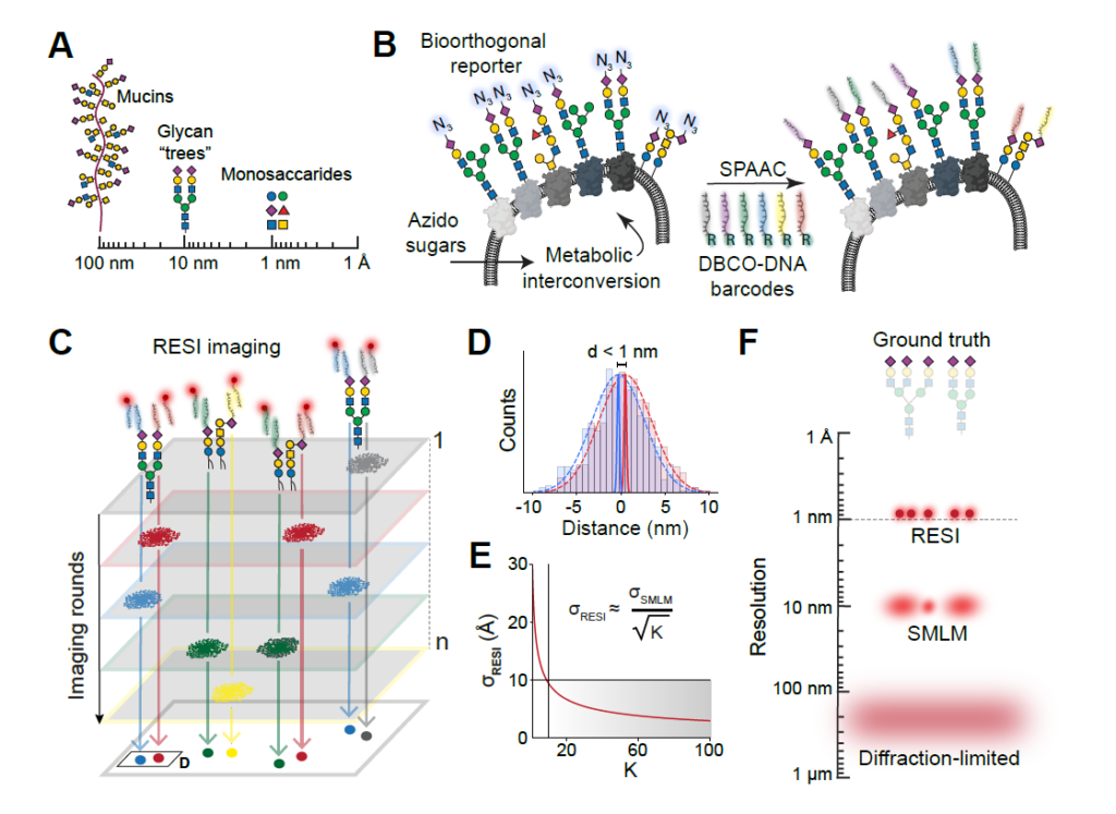
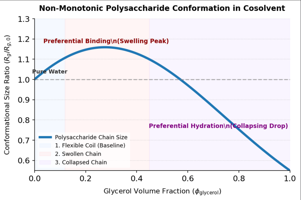
Fig. 1. Experimental concept. (A) Monosaccharides represent the smallest length scale in glycobiology, forming the fundamental building blocks for larger glycan structures (tens of nanometers) and heavily glycosylated mucins (up to hundreds of nanometers). Monosaccharides are depicted following the Symbol Nomenclature for Glycans (SNFG) guidelines (55). (B) Azido sugars are metabolized by cells and integrated into target monosaccharides, introducing a bioorthogonal azido group as a molecular reporter. This azido group facilitates the attachment of six orthogonal DBCO-modified DNA strands (shown in different colors) via a strain-promoted azide-alkyne click chemistry reaction (SPAAC), enabling precise labeling of the target monosaccharide unit. (C) Imaging sugar molecules, labeled with distinct DNA barcodes, through the sequential addition of their corresponding imaging DNA sequences, facilitates temporal separation of signals distinguishing blinks from nearby molecules. (D) Combining all localizations per target (𝐾) from each imaging round enhances localization precision. (E) In RESI, localization precision improves with 1⁄√𝐾, thus, resolution enhancement is independent of 𝜎!”#”, achieving Angstrom-scale precision. (F) Unlike other super-resolution imaging techniques, RESI can resolve single sugars within a glycan. The glycan structures exemplify sialic acid labeling.

Latest news

This study uncovers a surprising, non-linear behavior in how polysaccharides change shape when you add...

Isotopic metabolomics reveals that plant species exhibit highly divergent carbon allocation and metabolic rewiring strategies...

The Protein Data Bank (PDB), established in 1971, is the primary global, open-access archive for...

Carbohydrates are among the most abundant and structurally diverse biomolecules in nature, playing central roles...