The chemical and biological mechanisms leading to lignin assembly in the plant cell wall into a complex 3D structure are not elucidated. Little is known about its supramolecular organization. What makes lignin polymerization in planta a peculiar phenomenon is that lignification consists of the formation of a hydrophobic polymer within the already organized chiral and hydrophilic polysaccharide environment of the maturing cell wall. During plant development, lignin monomer biosynthetic genes are thought to be expressed in all lignifying tissues as well as in non-lignifying tissues, as inferred in the “good neighbor hypothesis” which proposes that lignin precursors are synthesized in nonlignified cells and exported to the walls of adjacent lignifying cells Farquharson 2013Smith et al., 2013.
It is not clear how lignin deposition is directed to specific sites of the cell walls. It has been proposed that NADPH oxidases and the reactive oxygen species (ROS) that they produce might provide specificity and subcellular precision in the way plant cells regulate lignin formation Li, Chapple, 2010. Early studies using radiolabeled monolignol precursors Takabe et al., 1985 TerashimaFukushima 1988Kaneda et al., 2008 allowed to identify lignification sites in the cell wall. Contrary to a long believed concept, lignin deposition is not exclusively a postmortem event, but occurs prior programmed cell death, as evidenced by immuno-gold labeling in transmission electron microscopy Ruel et al., 2006 and further demonstrated by the distribution of radioactively labeled phenylpropanoids Smith et al., 2013.
Recently, the use of fluorescence-tagged monolignols and click chemistry-assisted covalent labeling enabled imaging their incorporation into plant tissue, providing information about the localization of lignification during plant cell wall maturation and lignin matrix assembly Tobimatsu et al., 2013.
Computational Modeling
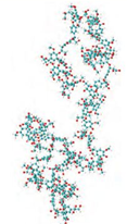
Since the early representations of lignin chemical formulas by Freudenberg (1968) or Adler (1977), several structures of lignins have been published. In spite of the significant analytical progress of the recent years, all of these representations are nothing more than average schematic images of the statistical occurrence of the main linkages found in lignins from various origins, but by no means do they provide a clear and accurate molecular structure of these complex polymers. Adding the stereochemical and conformational complexities associated with a molecular representation of even a model lignin containing 20 phenylpropanoid units, Ralph et al., 200) calculated that the 38 optical centers of such a molecule could generate 234 (i.e. 17,179,869,184) theoretical isomers ! A long time after the early computer model of lignin by Glasser and Glasser in 1974, more recent computational simulation studies have refined the stereochemical and conformational knowledge of lignin model compounds. Besombes and Mazeau Besombes & Mazeau, 2004, 2005 have provided molecular dynamics calculations from β-O-4 dimers up to a 20-units oligomer. A modeling of 60 guaiacyl units has been computerized Smith et al., 2016(Fig. 5).Their results of molecular modeling showed that in a bilayer of 124 dimers adsorbed around a cellulose wisker the first layer of several lignin aromatic units is in a parallel orientation at the interface. This was in keeping with the experimental observation of lignin aromatic rings on cellulose surface obtained by Atalla and Agarwal (1985) Attalla & Agarwal; 1985 in Raman spectroscopy. In a study of the outer epidermal cell walls of wheat stems using Raman scattering spectroscopy Cao et al., 2006 showed that xylan and the phenylpropane units of lignin tend to lie perpendicular perpendicular to the net orientation of cellulose.

The aggregation of lignin by self-assembly has been suggested to display fractal properties Achyuthan et al., 2010. Indeed, the analysis of images obtained using scanning tunneling microscopy showed that lignin had fractal organization suggesting that the polymer could have a regular structure Radotic et al., 2000Zhao et al., 2016. It has not been shown, yet, that this concept applied in vivo to protolignin.
