Cell surface polysaccharides in the gut microbiota: occurrence, structure and role

Author(s)

V. Laplanche, I. Speciale, C. De Castro & N. Juge

Sources

Victor Laplanche, Immacolata Speciale, Cristina De Castro & Nathalie Juge (2025) Cell surface polysaccharides in the gut microbiota: occurrence, structure and role, Gut Microbes, 17:1, 2536082, DOI: 10.1080/19490976.2025.2536082 https://doi.org/10.1080/19490976.2025.2536082

The gastrointestinal (GI) tract is home to trillions of microorganisms that live in symbiosis with their host. Commensal bacteria in the gut communicate with epithelial and immune cells via effector molecules that are either secreted or attached to the cell surface. Although cell surface polysaccharides have primarily been studied in the context of pathogen-host interactions, they are increasingly being recognized as essential factors in the symbiotic interaction between gut microbiota and host, conferring biological activities and physiological functions. This review focuses on the structure and function of polysaccharides surrounding the bacterial cell wall: capsular polysaccharides (CPS), which are tightly linked to the cell surface; cell wall polysaccharides (CWPS), which are also tightly linked to the cell surface; and exopolysaccharides (EPS), which are loosely attached to the extracellular surface or secreted into the environment. Our focus will be on structurally characterized CPS, CWPS, and EPS from gut commensal and food-derived bacteria. These polysaccharides exhibit significant structural diversity, enabling bacteria to adapt to the GI environment and/or influence the host’s immune response. The combined diversity of microbes in the gut provides a vast array of glycans that could be exploited for the benefit of human health.
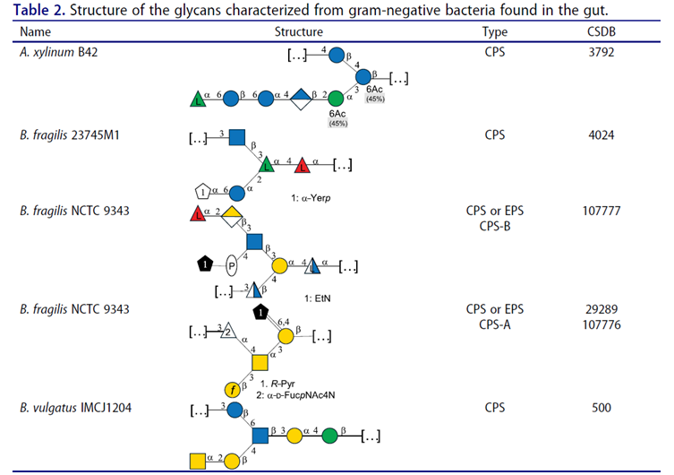
Figure  A.” stands for Acetobacter, and “B.” stands for Bacteroides, the Carbohydrate Structure Database (CSDB) identification number is given in the corresponding column. The structures are depicted according to the rules given by the Systematic Nomenclature of Glycans. Where not specified, all the residues are in the pyranose form (if furanose, a “f” is reported inside the symbol). All residues have the D absolute configuration unless otherwise specified.

Latest news

Oligosaccharyltransferase (OST) catalyses the key step of N-glycosylation, transferring immature N-glycans to select Asn...

Pectin can be divided into four distinct structural categories: homogalacturonan, xylogalacturonan, rhamnogalacturonan I (RG-I), and...

Glycans are complex carbohydrates that exhibit extraordinary structural complexity and stereochemical diversity while playing essential...

Efforts to fully understand the structure-to-function relationships of glycosaminoglycans (GAGs) have been hampered by the...