Structure of the Lipopolysaccharide from Paenalcaligenes hominis: A Chemical Perspective on Immune Recognition

Author(s)

F. Nieto-Fabregat, Q. Zhu, C. Vivès, Y. Zhan, A. Marseglia, F. Chiodo, M. Thépaut, D. Rai, S.S. Kulkarni, F. Di Lorenzo, A. Molinaro, R. Marchetti, F. Fieschi, G. Xiao, B.Yu & A. Silipo

Sources

JACS Au 2025, 5, 3311−3327

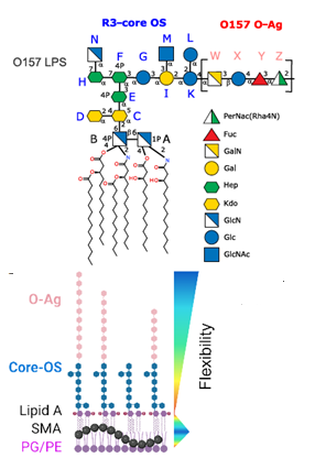
The Gram-negative bacterium Paenalcaligenes hominis is increasingly prevalent in elderly individuals and is associated with cognitive decline and dysfunction of the gut-brain axis. In this study, the authors present a comprehensive structural characterization of P. hominis lipopolysaccharide (LPS), which is a key modulator of immune recognition and the main component of the bacterium’s outer membrane. They unveil a unique O-antigen characterized by a trisaccharide repeating unit containing rhamnose and glucosamine. This O-antigen displays non-stoichiometric O-acetylation and has a terminal methylated rhamnose capping the saccharide chain. Furthermore, they disclose a short core oligosaccharide and a Lipid A composed of pentacyclic to tetracyclic species. Notably, this LPS exhibits reduced activation of Toll-like receptor-dependent signaling compared to highly immunostimulatory Escherichia coli LPS, and it elicits a poor pro-inflammatory cytokine response. Furthermore, P. hominis LPS exhibits selective binding to immune lectins, including Ficolin-3 and Galectin-4. This raises the possibility that lectin-mediated recognition may represent an alternative route of immune engagement that could explain the altered immune responses observed in elderly individuals. These findings lay the groundwork for further exploration of the role of P. hominis LPS in microbiota-induced immune modulation and its potential impact on age-related inflammatory and neurodegenerative conditions.

Figure: Computational study of the MD of P. hominis O-antigen and LPSOdeAc tetradecasaccharides. (A) Superposition of all poses obtained during the MD simulation of the O-antigen tetradecasaccharide. (B) Most representative pose of the O-antigen tetradecasaccharide (white), highlighting acetyl groups in teal. The terminal sugar (B residue) is highlighted in pink, with its terminal OMe group shown in cyan, displayed both with and without the molecular surface representation. (C) Most representative pose of LPSOdeAc tetradecasaccharide (green), also highlighting the terminal sugar (pink) and its OMe group (cyan), shown with and without surface. (D) Front view of O-antigen (white) and LPSOdeAc (green) tetradecasaccharides, emphasizing their similar helical conformations.

/

Latest news

The glycans found on the surface of all immune cells are the product of diverse...

Starch granules offer a unique platform for studying complex, multiresonator lasing phenomena, highlighting the importance...

Cyclodextrins (CDs) are known for their ability to form supramolecular interactions with a wide range...

Lipids are essential components of cancer cells due to their structural and signalling roles. To...