Imaging of biomolecules guides our understanding of their diverse structures and functions. Real-space imaging at sub-nanometre resolution using cryo-electron microscopy has provided key insights into proteins and their assemblies. Direct molecular imaging of glycans has not been possible so far. The inherent glycan complexity and backbone flexibility require single-molecule approaches for real-space imaging. At present, glycan characterization often relies on a combination of mass spectrometry and nuclear magnetic resonance imaging to provide insights into size, sequence, branching and connectivity, and therefore requires structure reconstruction from indirect information.
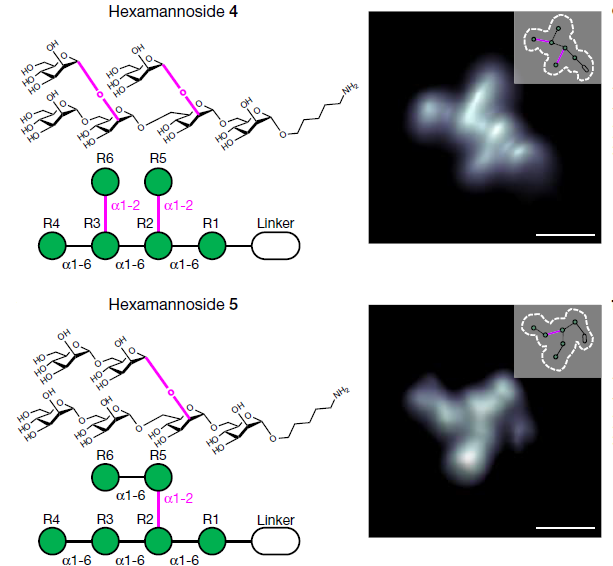
The authors present direct imaging of single glycan molecules that are isolated by mass-selective, soft-landing electrospray ion beam deposition and imaged by low-temperature scanning tunnelling microscopy. The sub-nanometre resolution of the technique enables the visualization of glycan connectivity and discrimination between regioisomers. Direct glycan imaging is an important step towards a better understanding of the structure of carbohydrates.